Research Products and Services
Covid-19 Proteomics related tools
We are committed to helping the global scientific community in their research and development endeavors to mitigate the COVID-19 pandemic by finding diagnostic and therapeutic solutions. We offer catalog peptides, fluorescent dyes, and assay kits. We also develop customized solutions.
With over 25 years of experience in serving the life science industry, we are ready to provide you with high quality, consistency, and safety.
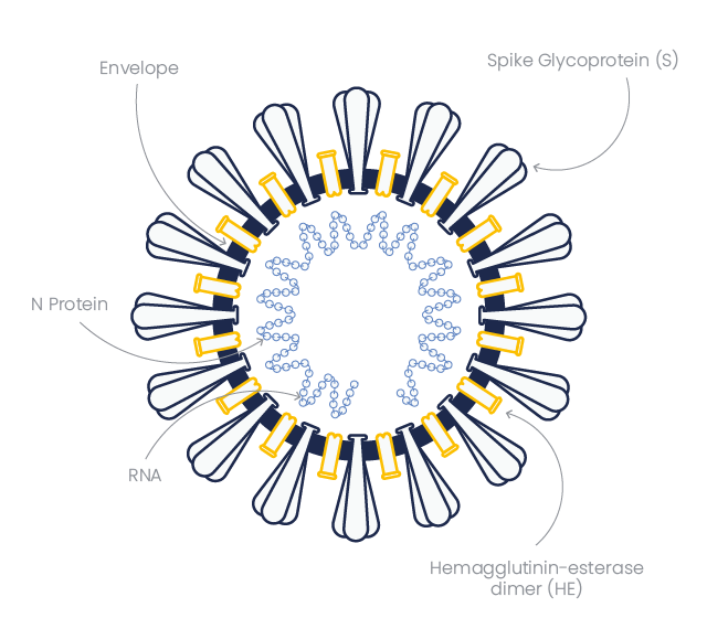
SARS-CoV coronaviruses
Severe acute respiratory syndrome coronavirus 2 (SARS-CoV-2, 2019-nCoV), the coronavirus responsible for coronavirus disease 2019 (COVID-19), shares very high similarity with SARS-CoV.
Both belong to the beta-coronaviruses family, which are enveloped, single-stranded RNA viruses. They mainly infect host lung cells through binding to the Angiotensin Converting Enzyme 2 (ACE2) receptor.
Like other coronaviruses, SARS-CoV-2 has four structural proteins: Spike glycoprotein (S), Nucleocapsid (N), Envelope (E), and Membrane (M) proteins.
Spike glycoprotein :
a diagnostic and therapeutic target
The Spike glycoprotein mediates the virus attachment to host cell surface receptors and facilitates virus entry
by assisting fusion between viral and host cell membranes. It is the most exposed and
immunogenic viral protein and hence a target of choice for diagnostic and therapeutic assays.
The Spike glycoprotein of SARS-CoV-2 has two furin-like protease cleavage sites. One of the sites is at the boundary between
S1 and S2 subunits having poly-basic residues, which is characteristic of SARS-CoV-2. The other cleavage site is located within the S2 subunit.

Schematic representation of the Spike protein of SARS-CoV and SARS-CoV-2:
SP: signal peptide; NTD: N-terminal domain; RBD: receptor binding domain; RBM: receptor binding motif; FP: fusion peptide;
HR1: heptad repeat 1; HR2: heptad repeat 2; TM: transmembrane domain; CD: cytoplasmic domain. The S1/S2 cleavage site is indicated.
Your provider for COVID proteomics


Protease Substrates
Peptides play various roles in the Coronavirus physiology. For example, certain sequences can serve as substrates for the proteolytic machinery of the viral fusion system, epitopes for antibody screening, targets within receptor binding or fusion domains or have inhibitory roles in viral autophagy.
| Product Name | Catalog # | Order |
| ACE2 Substrate | AS-60757 | Order |
| DX 600, ACE2 Inhibitor | AS-62337 | Order |
| Generic 3CLpro FRET peptide substrate NEW HilyteTM Fluor - 488 - ESATLQSGLRKAK - (QXL® - 520) - NH2 |
AS-65599 | Order |
| pro - NPY peptide (34 - 43), human NEW QXL®520 - RQRYGKRSSP - K(5 - FAM) - NH2 |
AS-65602 | Order |
| Spike protein (S1/S2) SARS - CoV - 2 substrate NEW QXL®520 - TNSPRRARSVAS - K(5 - FAM) - NH2 |
AS-65600 | Order |
| Spike protein (S2) SARS - CoV - 2 substrate NEW QXL®520 - SKPSKRSFIED - K(5 - FAM) - NH2 |
AS-65601 | Order |
| Tat-Beclin-1 | AS-65467 | Order |
| Tat-Beclin-1, scrambled | AS-65468 | Order |
Assay kits
The SARS-CoV viral proteins have been identified as targets of several host proteases, among which Furin,
3CLpro (3C-like viral protease) and Cathepsins (B, L) play roles.
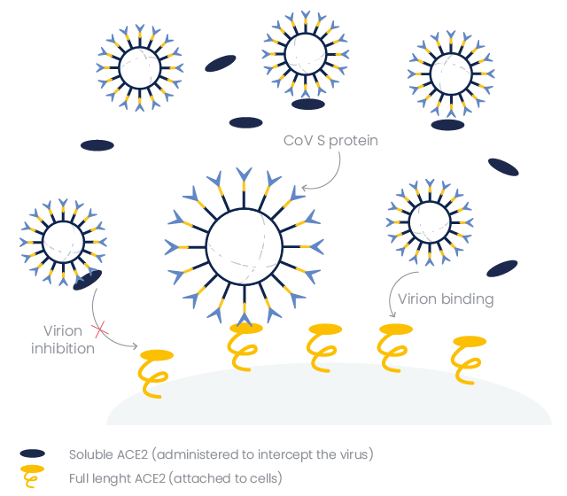
A recent study, Battle D. et al. (2020) have shown that a soluble form of ACE2 may act as a competitive inhibitor
of SARS-CoV-2 and other coronaviruses by preventing the binding of the viral particle to the membrane-bound full-length form.
AnaSpec (Eurogentec's US subsidiary), the expert in FRET peptide substrates, offers a range of catalog FRET peptides labeled
with our QXL® quenchers and fluorescent dyes. Custom sequences can be provided.
FRET peptide substrates are key components of our protease activity assay kits.
ACE2 Assay Kits
The extracellular domain of ACE2 has been demonstrated as a receptor for the spike (S) protein of SARS-CoV & SARS-CoV-2.
The soluble form of ACE2 may act as a competitive inhibitor of SARS-CoV-2 and could serve as a potentially novel therapeutic target to limit infection caused by SARS-CoV-2 (COVID-19).
Cited in high impact journals, Our FRET assay detects the activity of sub-nanogram level of ACE2
Cathepsin Assay Kits
Certain viruses including the Coronaviruses depend on cathepsins (L & B) for entry into their target cells.
The cleavage by proteases, renders them active for fusion with the host cell membrane.
Such assays can help identify and screen for inhibitors that can prevent cathepsin cleavage of viral glycoproteins.
Furin Assay Kits
The spike protein of some coronaviruses is cleaved at the S1/S2 boundary by furin(-like) proteases during transport of the newly assembled virions through the secretory pathway. Hence, screening for furin inhibitors observed with furin-dependent viral replication may present as a potential target for drug design.
Featured Citations
Measurement of Angiotensin Converting Enzyme 2 Activity in Biological Fluid (ACE2)
Xiao F, Burns K.D. (2017)
In: Touyz R., Schiffrin E. (eds) Hypertension. Methods in Molecular Biology, vol 1527. Humana Press, New York, NY.
Identification of a broad-spectrum antiviral small molecule against severe acute respiratory syndrome coronavirus and Ebola, Hendra, and Nipah viruses by using a novel high-throughput screening assay.
Elshabrawy HA, Fan J, Haddad CS, et al.
J Virol. 2014;88(8):4353–4365.
SKP2 attenuates autophagy through Beclin1-ubiquitination and its inhibition reduces MERS-Coronavirus infection.
Gassen NC, Niemeyer D, Muth D, Corman VM, Martinelli S, Gassen A, Hafner K, Papies J, Mösbauer K, Zellner A, Zannas AS.
Nature Communications. 2019 Dec 18;10(1):1-6.
Ligand-induced Dimerization of Middle East Respiratory Syndrome (MERS) Coronavirus nsp5 Protease (3CLpro) IMPLICATIONS FOR nsp5 REGULATION AND THE DEVELOPMENT OF ANTIVIRALS.
Tomar S, Johnston ML, John SE, Osswald HL, Nyalapatla PR, Paul LN, Ghosh AK, Denison MR, Mesecar AD.
Journal of Biological Chemistry. 2015 Aug 7;290(32):19403-22.
Dissecting virus entry: replication-independent analysis of virus binding, internalization, and penetration using minimal complementation of β-galactosidase.
d C, Bloyet LM, Wicht O, et al.PLoS One. 2014;9(7):e101762.
References
- 1. Li, W. et al. (2003) Nature 426, 450.
- 2. Lu R., et al. (2020) Lancet 395, 565–574
- 3. Du, L., et al. (2009) Nat. Rev. Microbiol 7, 226–236
- 4. Elshabrawy HA, et al. (2014). Journal of Virology. 88(8), 4353-65.
- 5. Burkard C, et al. (2014). PLoS One. 2014; 9(7):e101762.